
Updated 03/29/24
Retinacula are essential components of the musculoskeletal system, particularly in the distal extremities, where they facilitate efficient and smooth movement. These fibrous bands or thickenings of the deep fascia hold tendons in place near joints, acting as pulleys to maintain the mechanical efficiency of the musculotendinous units. This discussion will delve into the biomechanics, anatomy, and clinical implications of retinacula, focusing on their role in the distal extremities and their relevance to massage and manual therapy.
Biomechanics and Anatomy:
Retinacula are most prominent in the distal portions of the limbs, where long tendons must navigate sharp angles to reach their insertions. The extensor retinaculum of the ankle and the flexor retinaculum of the wrist are prime examples. These structures hold the tendons close to the joint, preventing them from bowstringing away during muscle contraction. This pulley-like mechanism ensures that the force generated by the muscle is efficiently transferred to the tendon’s insertion point, allowing for smooth and precise movement.
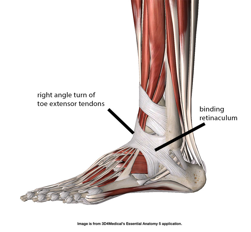
The muscles of the distal extremities are much different. In the long tendons of the hands and feet the tendons do not have a straight line of pull. In some instances the tendon must take almost a right angle turn. The long extensors of the toes are a good example. They travel the length of the lower leg and then over the top of the foot to the toes. They take close to a 900 angle bend over the top of the foot near the ankle joint (Image 1).
The retinacula are composed of dense connective tissue, primarily collagen fibers, which provide strength and stability. They are attached to the underlying bone and blend with the deep fascia of the surrounding muscles. The retinacula are relatively avascular, receiving their blood supply from the adjacent periosteum and synovial sheaths.
Innervation and Pain Conditions:
The retinacula around the distal extremities are not extensively innervated, but they are near various neurovascular structures. For example, the flexor retinaculum of the wrist forms the roof of the carpal tunnel, which contains the median nerve and flexor tendons. Compression or irritation of these structures can lead to conditions such as carpal tunnel syndrome, characterized by pain, numbness, and weakness in the hand and wrist.
Similarly, the ankle flexor retinaculum is closely related to the tibial nerve and its branches, which can be compressed or irritated, leading to conditions like tarsal tunnel syndrome. This condition can cause pain, numbness, and weakness on the plantar surface of the foot and is often mistaken for plantar fasciitis.
As previously discussed, Tenosynovitis is another common pain condition associated with retinacula. The high pressure between the tendons and their restraining retinacula can lead to irritation and inflammation of the synovial sheaths, causing pain and restricted movement. A prime example is “Lace bite,” a tenosynovitis of the extensor tendons at the ankle.
Massage and Manual Therapy Considerations:
When addressing pain conditions related to retinacula, massage therapists should consider the delicate balance between mobilizing the affected tissues and avoiding further irritation. Gentle friction massage along the length of the retinaculum may help reduce adhesions and improve the tendons’ gliding. However, caution should be exercised, as excessive pressure or friction can exacerbate the irritation of the synovial sheaths.
In conditions like carpal tunnel or tarsal tunnel syndromes, massage therapy can be beneficial in reducing muscle tension and promoting tissue mobility in the affected area. Techniques such as myofascial release, gentle stretching, and nerve gliding exercises may help to alleviate symptoms and improve overall function.
The retinacula plays a vital role in the biomechanics of the distal extremities, acting as pulleys to facilitate efficient and smooth movement. However, their close relationship with neurovascular structures and their high pressures on tendons can also contribute to various pain conditions. As massage therapists, understanding the anatomy and function of retinacula is crucial for effectively addressing these conditions through manual therapy techniques while recognizing the importance of a multidisciplinary approach to patient care.