Understanding Vertebrobasilar Insufficiency
- Whitney Lowe
Tightness and trigger points in the cervical muscles, and especially the sub-occipital muscles, often cause muscle tension headaches, so these are vital muscles to address in many of our massage treatments. There are a number of ways to access the cervical muscles, but there are also certain essential precautions when treating the neck. One of these concerns involves potentional artery vulnerability in the sub-occipital region. There are two key arteries in the suboccipital region susceptible to adverse compression. The compression of these arteries produces a condition called vertebrobasilar insufficiency (VBI), which can be dangerous. Let’s take a look at the structure and mechanics of the suboccipital region to understand how this condition occurs and how to avoid these complications in our treatments.
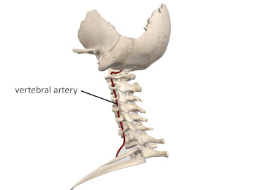
There is a vertebral artery on each side of the cervical region. The vertebral arteries on each side branch off from the subclavian artery at the base of the neck. After leaving the subclavian artery, they course through a small opening in the transverse processes of each cervical vertebrae called the transverse foramen (plural: foramina) (Figure 1).

Figure 1
Vertebral arteries passing through transverse processes in cervical vertebrae
Image is from 3D4Medical’s Complete Anatomy application
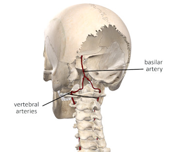
After passing through the transverse foramen in the C1 vertebra, the left and right vertebral arteries join together to form the basilar artery as they enter the foramen magnum of the skull and supply blood to the brain (Figure 2).

Figure 2
Vertebral arteries joining together to form basilar artery
Image is from 3D4Medical’s Complete Anatomy application
VBI is a condition caused by compression of either vertebral artery or compression of the basilar artery (after the two have joined together). The primary adverse impact of VBI is a lack of blood flow to the brain from arterial compression. Depending on each individual’s unique anatomical structure, the space between the occiput and the C1 vertebra (atlas), may be quite small. When the head moves into extension or rotation, these positions may further decrease the space between the occiput and C1 vertebra and compress one or both vertebral arteries. While extension and rotation head movements are the primary culprits that produce VBI, direct compression of the arteries from manual therapy techniques applied to the suboccipital region can also create VBI.
Other factors can cause VBI such as nearby bone spurs, small tumors, or plaque buildup within the vertebral arteries. If some other structure is leading to partial compression of the arteries, a thrombus (clot) is even more likely to develop from within the affected arteries. Sometimes a small slip of tendon attachment from muscles attaching to the cervical vertebrae may be close enough to the vertebral arteries to produce the adverse compression. When taking a client history, be aware that other common risk factors for cardiovascular disease such as smoking, hypertension, age, genetics, and family history may also increase the risk for VBI.
Impaired blood flow from VBI may cause dizziness, blurred vision, ringing in the ears, vertigo, or fainting in more severe cases. In some instances compression of one vertebral artery may be tolerable because blood can get to the brain through the other. However, if both vertebral arteries are compressed or the compression on these structures is higher up on the basilar artery, then there is a higher risk of an adverse event.
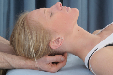
A common technique in massage that could potentially cause problems with VBI is when the practitioner applies bilateral compression to the client’s suboccipital region simultaneously with the fingertips (Figure 3). This fingertip pressure applied to both vertebral arteries simultaneously could obstruct blood flow sufficiently to produce VBI. If the head is in some degree of extension or rotation at the same time as this technique, there is an even greater risk of vertebral artery compression. An alternative to having the fingers of both hands pressing into the sub-occipital region (and possibly compressing both vertebral arteries) is to have only one hand doing this at a time.

Figure 3
Bilateral compression to the vertebral arteries
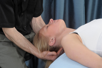
Another potential problem is any technique that involves significant hyperextension of the head such as a stretching treatment for the anterior cervical muscles (Figure 4). Hyperextension decreases the suboccipital space and may compress either the vertebral arteries or the conjoined basilar artery.

Figure 4
Vertebral artery compression from head hyperextension
The client may report symptoms of vertebrobasilar insufficiency within a short time if a particular position or technique compresses the arteries. However, they may also appear relaxed and you may not be aware they are having any of these symptoms, so be sure to check in with them frequently about any unusual sensations they are having when performing these techniques. If any technique causes vertebrobasilar insufficiency, immediately stop that technique, remove any pressure and bring the person back into a neutral head position. Be watchful for these signs or symptoms because they can come on quickly without notice.
VBI is not a common condition, and in most cases you can perform suboccipital compression or head hyperextension techniques without any adverse effects. However, if VBI occurs, the outcomes could be quite serious, so it is vital to be aware of these potential problems.

